September 2014 – Using viral transcription profiling, this study demonstrates the existence of multiple subtypes of Kaposi Sarcoma lesions for the first time in HIV- and Kaposi Sarcoma-treatment-naive patients.
Viral Profiling Identifies Multiple Subtypes of Kaposi’s Sarcoma
Mina C. Hosseinipour, Kristen M. Sweet, Jie Xiong, Dan Namarika, Albert Mwafongo, Michael Nyirenda, Loreen Chiwoko, Deborah Kamwendo, Irving Hoffman, Jeannette Lee, Sam Phiri, Wolfgang Vahrson, Blossom Damania, Dirk P. Dittmerd
mBio
Full text available at PubMed Central and mBio
Abstract
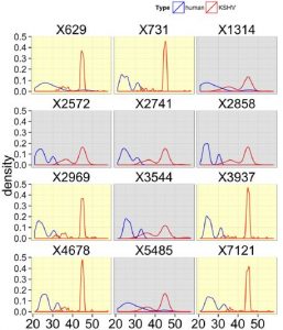
Kaposi’s sarcoma (KS), caused by KS-associated herpesvirus (KSHV), is the most common cancer among HIV-infected patients in Malawi and in the United States today. In Malawi, KSHV is endemic. We conducted a cross-sectional study of patients with HIV infection and KS with no history of chemo- or antiretroviral therapy (ART). Seventy patients were enrolled. Eighty-one percent had T1 (advanced) KS. Median CD4 and HIV RNA levels were 181 cells/mm3 and 138,641 copies/ml, respectively. We had complete information and suitable plasma and biopsy samples for 66 patients. For 59/66 (89%) patients, a detectable KSHV load was found in plasma (median, 2,291 copies/ml; interquartile range [IQR], 741 to 5,623). We utilized a novel KSHV real-time quantitative PCR (qPCR) array with multiple primers per open reading frame to examine KSHV transcription. Seventeen samples exhibited only minimal levels of KSHV mRNAs, presumably due to the limited number of infected cells. For all other biopsy samples, the viral latency locus (LANA, vCyc, vFLIP, kaposin, and microRNAs [miRNAs]) was transcribed abundantly, as was K15 mRNA. We could identify two subtypes of treatment-naive KS: lesions that transcribed viral RNAs across the length of the viral genome and lesions that displayed only limited transcription restricted to the latency locus. This finding demonstrates for the first time the existence of multiple subtypes of KS lesions in HIV- and KS-treatment naive patients.